Cells along the outer edge of the retina begin to swell and multiply before memory problems appear.
That subtle shift, visible only under specialized imaging, may signal the earliest stages of Alzheimer’s disease. Researchers at Houston Methodist report that these peripheral eye changes emerge before major failures occur in the brain’s waste-removal system, suggesting the eye could reveal risk years earlier than current diagnostics.
The study, led by biomedical engineer Stephen Wong, Ph.D., examined how retinal support cells behave during the first phase of disease development in mice genetically engineered to develop Alzheimer’s pathology. Findings appear online in the Journal of Alzheimer’s Disease.
“The eyes are indeed a window into the brain, but our study reveals that we have been looking at the wrong part of the window,” Wong said. “While most clinical eye exams focus on the central retina, the most critical early indicators of AD appear to be hidden at the periphery of the eye.”

Most eye exams concentrate on the macula, the central region responsible for sharp vision. The new work points instead to the peripheral retina, an area with a higher density of glial support cells.
Those cells, called Müller glia, help maintain balance in retinal tissue and support blood vessels. The research team wanted to understand how they change during the earliest disease stages.
“Since the peripheral retina contains more glial cells than the central retina, we wanted to understand how these types of cells and blood vessels interact in different parts of the retina in early-stage AD,” said first author Glori Das, a graduate research assistant in Wong’s laboratory and an M.D.-Ph.D. student at Texas A&M School of Medicine.
Experiments focused on 3-month-old female mice carrying Alzheimer’s-related mutations, compared with healthy controls. Scientists used fluorescent markers to examine proteins tied to glial function and waste clearance.
One protein drew particular attention: Aquaporin-4, which helps move fluid and metabolic waste through the nervous system. In Alzheimer’s brains, disruptions in this process contribute to buildup of amyloid-beta, a hallmark protein linked to the disease.
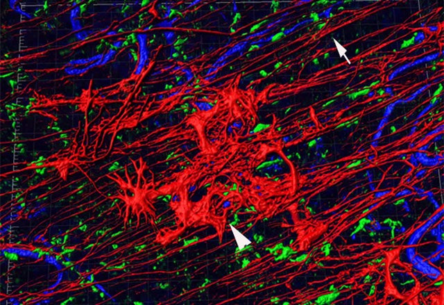
In the retina, Aquaporin-4 levels increased across all layers in the Alzheimer’s mice, with especially strong signals around blood vessels in the peripheral region. The mice also showed elevated levels of glial fibrillary acidic protein, an indicator that Müller cells were activated and under stress.
Researchers observed larger and more numerous glial cells in the retinal periphery. Wong described this as visual evidence that the body is working harder to maintain balance before breakdown occurs later.
Tracer experiments offered an unexpected result. Despite the cellular changes, overall fluid clearance along the optic nerve did not differ significantly between diseased and healthy mice at this stage. That finding suggests early retinal amyloid buildup may stem from increased local production rather than immediate failure of the clearance system.
The authors note that later dysfunction cannot be ruled out.
The work remains preliminary. It relies on mouse models, and the sample size was small, with five animals per group. Human studies will be necessary to confirm whether similar retinal changes occur before cognitive symptoms.
Still, the findings highlight a potential diagnostic advantage. Unlike brain imaging or spinal fluid testing, retinal imaging can be quick and noninvasive.
Wong said wide-field retinal scans could someday become routine in older adults, allowing doctors to detect Alzheimer’s risk during standard eye exams rather than through expensive procedures.
The research also suggests a new therapeutic target. If glial cell changes reflect early disease stress, treatments aimed at stabilizing these cells or improving fluid transport might slow progression.
Other authors on the study include Houston Methodist researchers Raksha Raghunathan, Lin Wang, Zhihao Wan, Matthew Vasquez, and Hong Zhao, who served as co-corresponding author. The work was supported by the T. T. and W. F. Chao Foundation.
Research findings are available online in the Journal of Alzheimer’s Disease.
The original story “The eyes may be a window into early Alzheimer’s detection, study finds” is published in The Brighter Side of News.
Like these kind of feel good stories? Get The Brighter Side of News’ newsletter.
The post The eyes may be a window into early Alzheimer’s detection, study finds appeared first on The Brighter Side of News.

