Cancer remains one of the world’s most serious health threats, especially when it spreads beyond its original site. That spread, known as metastasis, causes most cancer-related deaths. Now, researchers from the Hebrew University of Jerusalem report a new way to spot the most dangerous cancer cells by watching how they physically behave, rather than what genes or chemicals they carry.
The study introduces a fast, label-free method that distinguishes aggressive cancer cells by how they grip, stretch, and interact with their surroundings. The work was led by PhD student Chalom Zemmour under the guidance of Prof. Ofra Benny from the School of Pharmacy at the Hebrew University.
Instead of focusing on molecular markers, the team looked at something simpler and often overlooked. They asked how cancer cells respond when placed on a physically challenging surface. The answer, they found, can reveal how aggressive a cancer cell truly is.
For years, cancer cells have been classified using genetic tests and chemical signals. These tools are powerful, but they have limits. They can be costly, slow, and sometimes miss how threatening a cell actually is. Under a microscope, aggressive and less aggressive cancer cells can look nearly identical when grown on smooth laboratory dishes.
The new approach turns that problem on its head. Rather than studying what cancer cells express, the researchers focused on what cancer cells do.
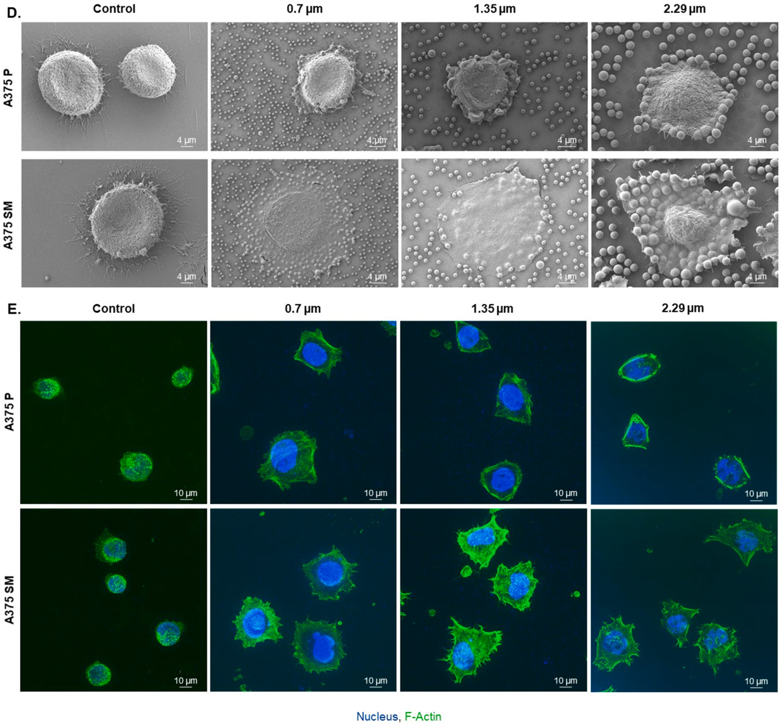
To make this possible, the team designed microscopic surfaces covered with tiny plastic beads. These beads form patterns at the nano and micro scale, far smaller than a grain of sand. To the naked eye, the surface looks smooth. To a cell, it feels like a rugged landscape.
When cancer cells land on these surfaces, they must push, pull, and adapt. That struggle reveals their true nature.
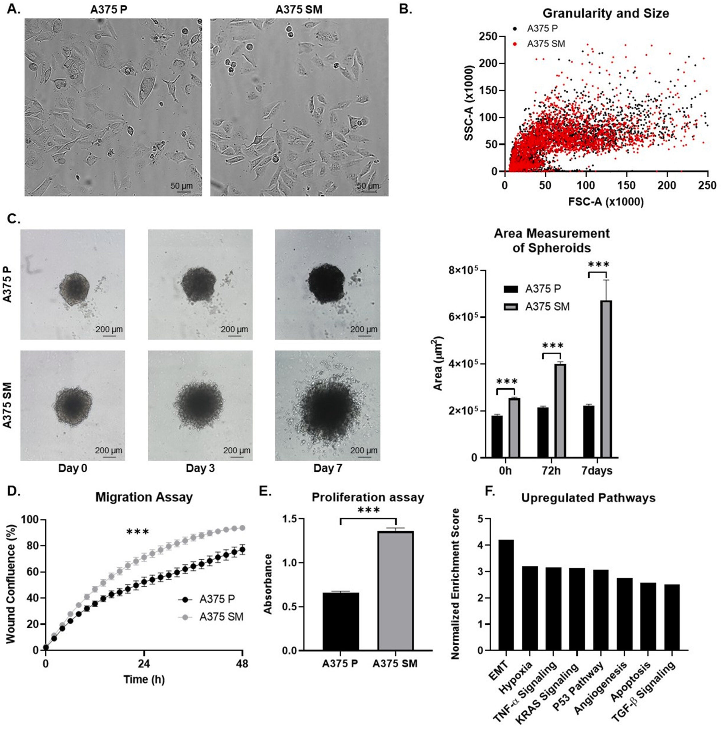
On the patterned surfaces, aggressive cancer cells behaved very differently from less harmful ones. The most dangerous cells gripped the surface tightly. They stretched their bodies and wrapped themselves around the tiny features. Many also swallowed more of the microscopic particles.
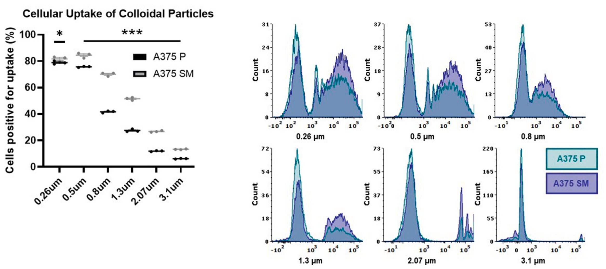
Less aggressive cells acted more cautiously. They attached weakly, showed limited stretching, and interacted far less with the surface. On standard flat lab plates, these differences almost disappear. The textured surface makes them impossible to miss.
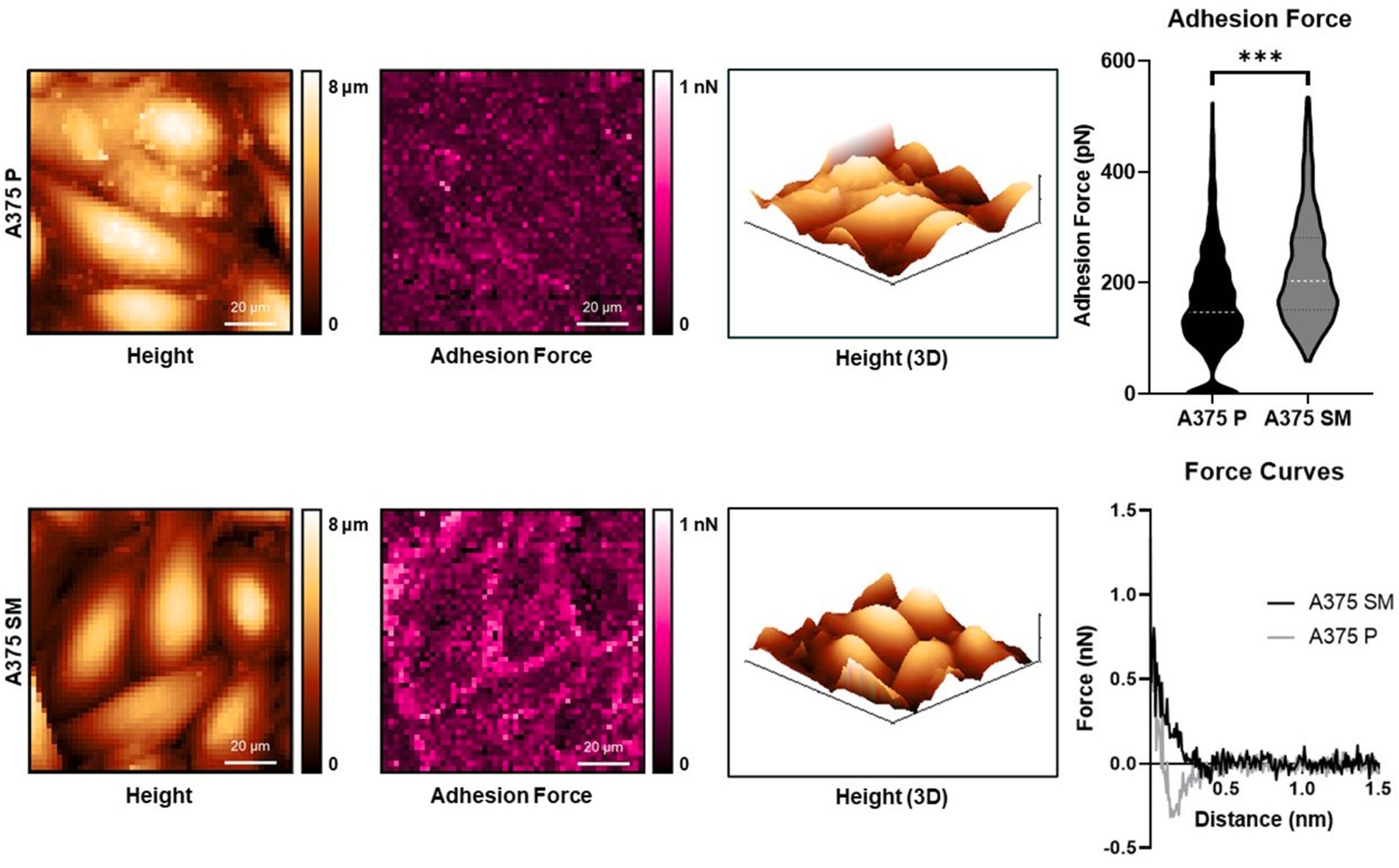
“This tells us that aggressiveness is not a fixed trait and we can have a sensitive technology to measure it,” Prof. Benny said. “It’s a functional state that can be revealed through physical behavior, not just molecular signatures.”
The surface acts like a mechanical sensor. It does not measure chemicals or genes. Instead, it reads how much force and effort a cell uses when faced with a physical challenge.
The study also offered new insight into metastasis itself. Cancer cells do not behave the same way at every stage of their journey through the body. The researchers found that cells change their mechanical behavior as they spread.
When cancer cells leave the original tumor, they often reduce how strongly they stick to surfaces. This loss of grip may help them travel through blood or tissue. Once they reach a new site, however, they regain strong adhesion and mechanical activity.
The patterned surfaces were sensitive enough to detect these shifts. That finding suggests cancer aggressiveness can rise and fall depending on where a cell is in the metastatic process.
Understanding these transitions could help researchers better predict when cancer is most likely to spread and when it might settle into new tissue.
One of the most striking aspects of this technology is how simple it is. The method does not require dyes, fluorescent labels, or genetic sequencing. The surfaces can be made using standard laboratory techniques and work with imaging tools already common in research labs.
Because of this, the approach could be adapted for many uses. It may help researchers quickly screen cancer cells for aggressive behavior. It could also support studies of tumor progression and metastasis. Drug developers might use it to test whether new treatments reduce the physical strength of cancer cells, not just their growth.
The technique may even support personalized cancer care. In the future, cells taken from a patient’s tumor could be placed on these surfaces to assess how aggressive they are. That information could help guide treatment decisions or monitoring strategies.
This work reflects a broader shift in cancer research. Scientists are increasingly recognizing that physical traits matter, not just molecular ones. How cells push, pull, move, and adapt can reveal risks that genes alone do not show.
“Our work shows that how cancer cells push, pull, and grip their surroundings can tell us a great deal about how dangerous they are,” Prof. Benny said. “This opens a new path for cancer diagnostics that is both powerful and surprisingly simple.”
While the technology is still in the research stage, its promise lies in accessibility. Because it builds on tools many labs already use, it could spread quickly if further studies confirm its value.
Cancer remains complex, and no single test will provide all the answers. Still, this approach adds a new and important perspective. It reminds researchers and clinicians that cancer is not just a chemical problem, but also a physical one.
This research could change how aggressive cancer is identified and studied. By offering a fast and label-free method, it may reduce reliance on costly genetic testing alone. It could help researchers better understand metastasis and how cancer cells adapt during spread.
In the future, the technology may support earlier detection of high-risk tumors, improve drug testing, and guide personalized treatment decisions. By focusing on how cells behave, not just what they contain, this work may help doctors identify danger sooner and respond more effectively.
Research findings are available online in the journal ScienceDirect.
The original story “Researchers find a new way to spot the most dangerous cancer cells” is published in The Brighter Side of News.
Like these kind of feel good stories? Get The Brighter Side of News’ newsletter.
The post Researchers find a new way to spot the most dangerous cancer cells appeared first on The Brighter Side of News.

