A small adhesive patch may soon give doctors a far clearer view of fetal health during pregnancy. Researchers at Stanford Medicine, the University of California San Diego and the University of Oxford have developed a wearable ultrasound patch that continuously tracks blood flow between a fetus and the placenta in real time.
The device could transform care for high-risk pregnancies, especially for conditions linked to poor blood flow and slow fetal growth. Unlike standard ultrasound exams that provide only brief snapshots, the new patch delivers continuous information while attached to the mother’s abdomen.
The research involved testing the device in dozens of pregnant women. Scientists say the technology could help physicians detect serious complications earlier and make faster decisions when fetal health begins to decline.
“There’s nothing similar to our device on the market or in the scientific literature,” said senior study author Sheng Xu, PhD, professor of anesthesiology, perioperative and pain medicine at Stanford Medicine.
Pregnancy complications tied to poor blood flow affect millions of families worldwide. One major condition, called intrauterine growth restriction, affects about 10% of pregnancies. In these cases, babies grow too slowly because they do not receive enough oxygen or nutrients through the placenta and umbilical cord.
Doctors currently rely on Doppler ultrasound scans and cardiotocography to monitor fetal health. Doppler ultrasound measures blood flow through vessels such as the umbilical artery, while cardiotocography tracks fetal heart rate and uterine contractions.
But both methods have limitations.
Traditional Doppler ultrasound requires a trained sonographer and scheduled appointments. The scans only capture short moments in time. Important changes between appointments may go unnoticed.
Cardiotocography can also be difficult to use continuously. Fetal movement often disrupts the signal, forcing nurses and physicians to reposition the monitor repeatedly.
“It’s really hard to be on that continuously,” said Jane Chueh, MD, a high-risk obstetrician at Stanford Medicine. “Even for inpatients, obtaining accurate readings three times a day can be labor intensive.”
These interruptions create stress for both families and medical staff. Physicians must constantly balance two dangerous possibilities: delivering a baby too early or waiting too long while blood flow worsens.
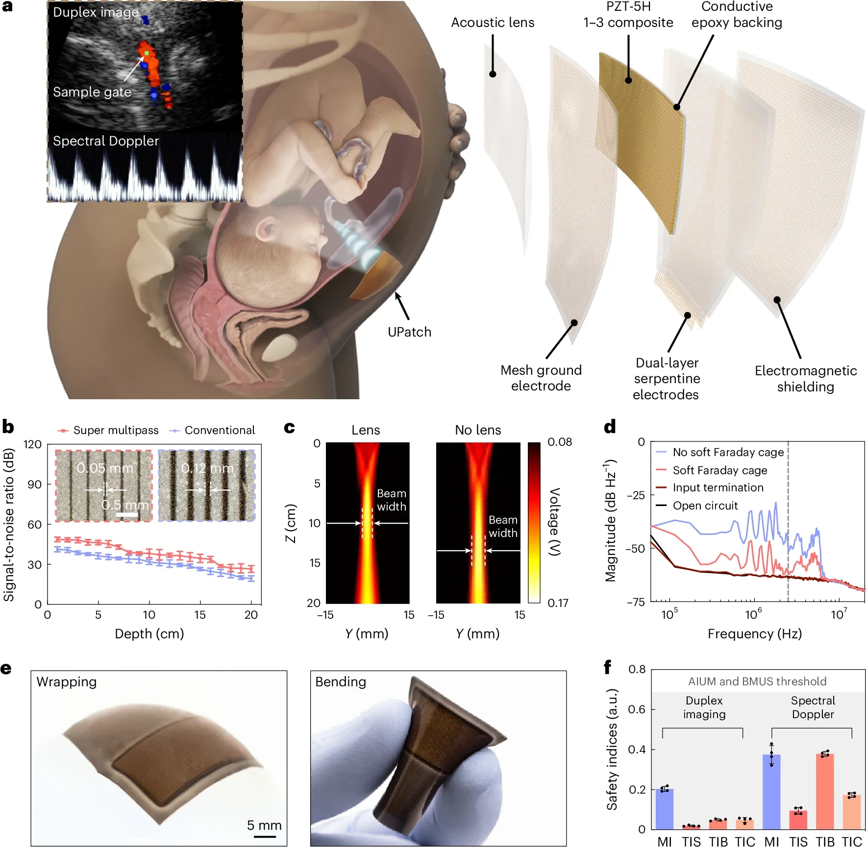
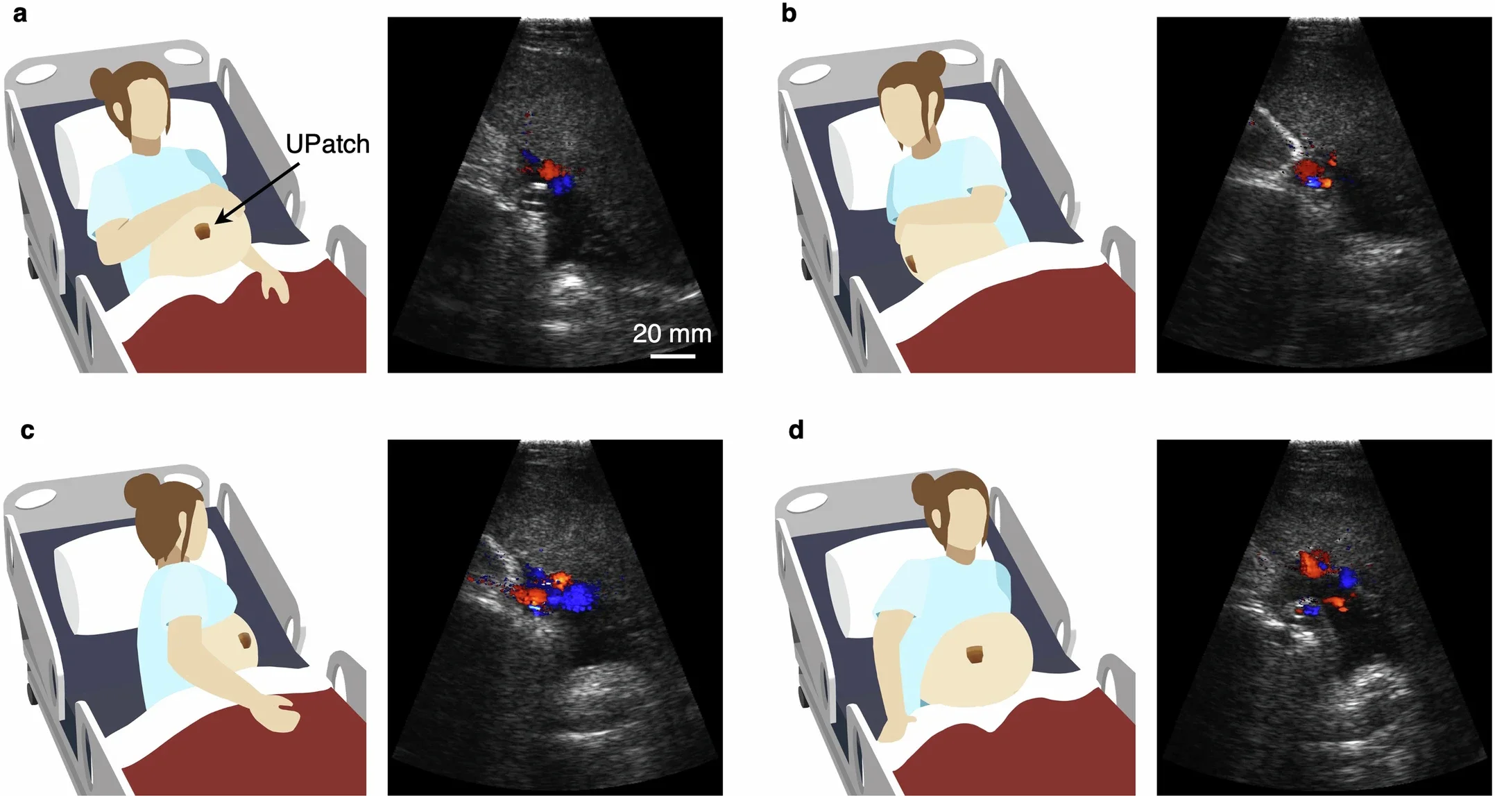
The new device, called the UPatch, aims to solve those problems. Roughly the size of a hand, the flexible patch adheres directly to the pregnant patient’s abdomen. It connects through a cable to a computer that processes ultrasound signals in real time.
Unlike handheld ultrasound probes, the patch does not require a sonographer to constantly adjust positioning. Instead, it uses built-in imaging algorithms to automatically track blood vessels and fetal structures even when the fetus or mother moves.
This presented a major engineering challenge.
The fetus changes position frequently inside the uterus. The umbilical cord floats freely in amniotic fluid. Traditional ultrasound systems depend on human operators who reposition probes constantly to maintain a clear image.
Researchers solved this problem by targeting the region where the umbilical cord connects to the placenta. That area remains relatively stable compared to the rest of the cord.
“We thought, ‘What if we target the ultrasound device onto the placenta, in the area where the umbilical cord attaches?’” said lead author Geonho “Tom” Park, PhD. “Even though everything is moving, there is some stability in the umbilical cord at that location.”
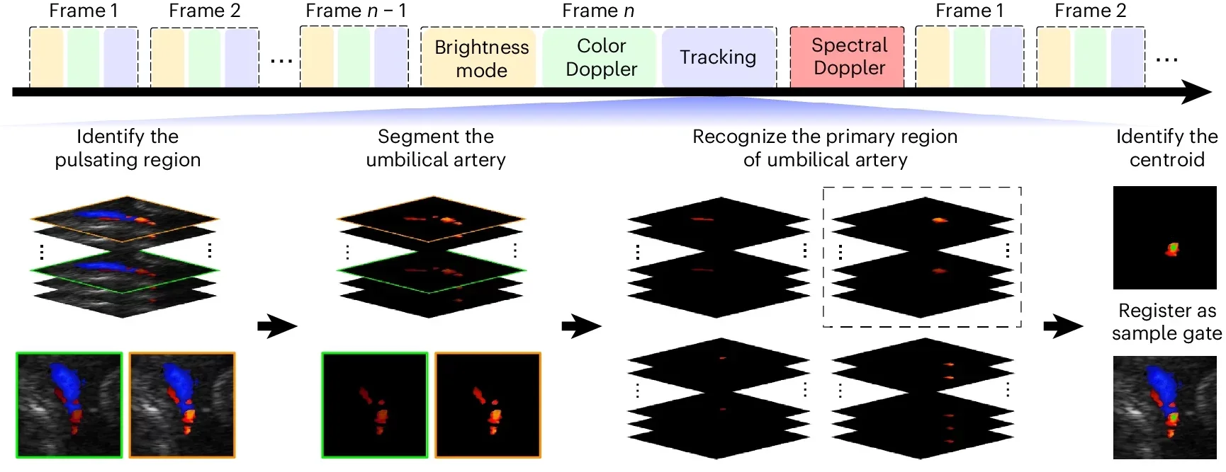
The team also developed a segmentation-based tracking algorithm that automatically follows blood vessels in real time. The software continuously identifies pulsating regions in Doppler images and adjusts the measurement window without human intervention.
Monitoring fetal circulation continuously requires detecting weak blood flow signals deep within the uterus. To achieve this, researchers redesigned several parts of the ultrasound system.
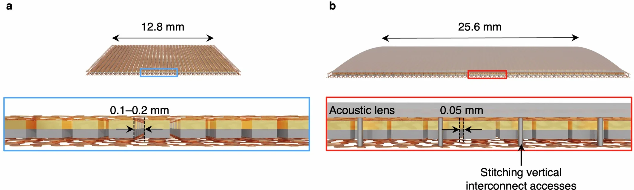
They created specialized ultrasound transducers using a fabrication process called super multipass dicing. This increased signal strength while reducing interference. The team also added an acoustic lens that sharpened image resolution and shifted the focal depth closer to the typical depth of the uterus.
A flexible Faraday shielding layer reduced electromagnetic interference around the device. Together, these changes improved the signal-to-noise ratio while keeping the patch flexible and comfortable.
Researchers also tested safety extensively. The patch operated well below ultrasound safety limits established by the U.S. Food and Drug Administration and major ultrasound societies.
Even after 48 hours of operation, the patch raised skin temperature by less than 0.7 degrees Celsius.
To validate the system, researchers tested the patch on 62 pregnant women and compared its results with standard clinical ultrasound machines.
The findings closely matched.
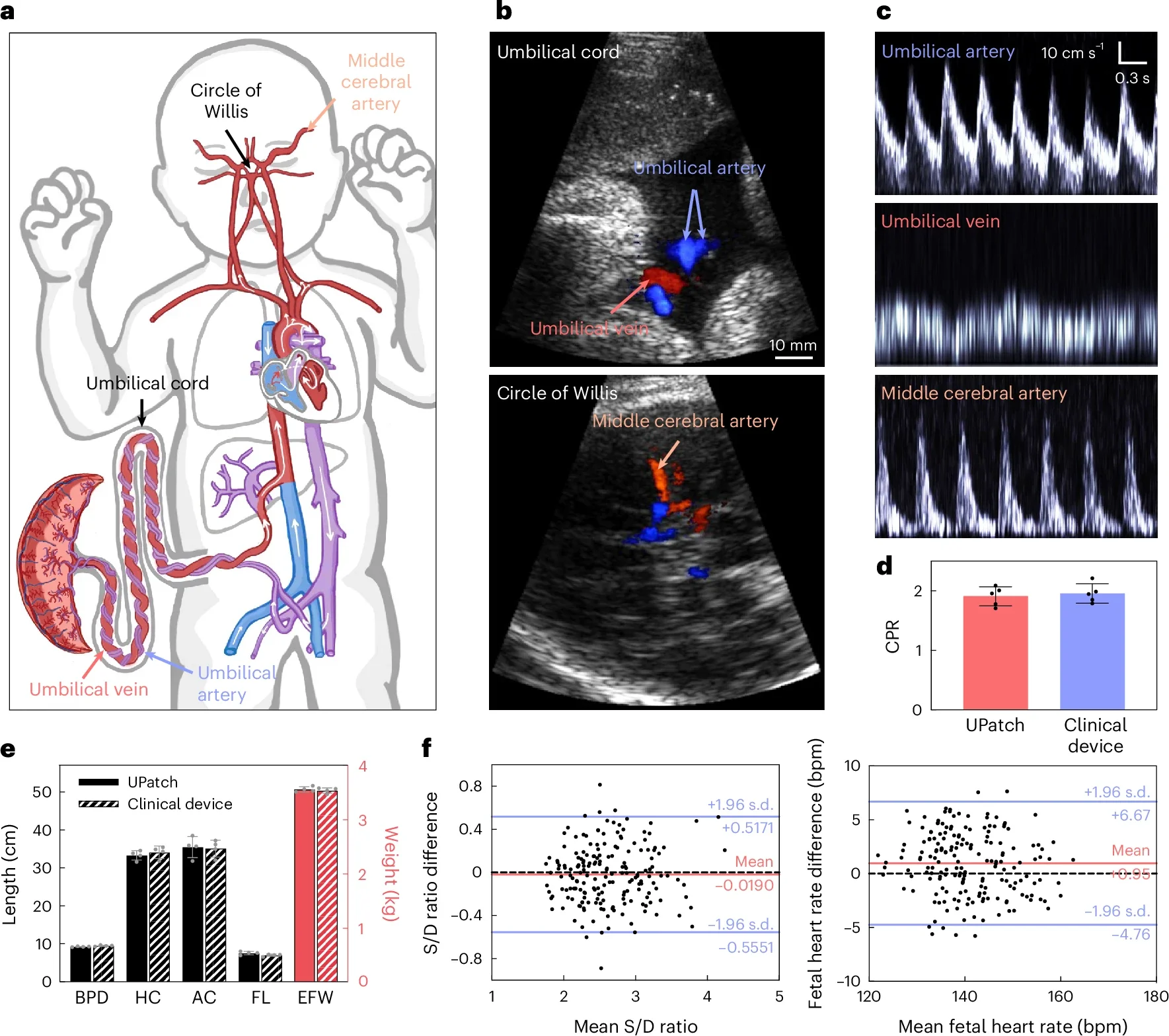
The patch accurately measured blood flow in the umbilical arteries and vein, as well as the fetal middle cerebral artery. It also measured fetal anatomy, including head circumference, abdominal circumference and femur length, which physicians use to estimate fetal weight.
Statistical comparisons showed extremely close agreement between the wearable device and standard ultrasound systems. Differences in blood flow measurements and fetal heart rate remained minimal.
The tracking algorithm also performed strongly. In more than 90% of tested images, the automated sample placement closely matched that of experienced sonographers.
Importantly, the system worked regardless of where the placenta was located inside the uterus.
During testing, the device unexpectedly identified a serious complication in one participant.
The patient was 28 weeks pregnant. Her fetal heart rate initially appeared normal. But the patch revealed major abnormalities in umbilical cord blood flow.
Park initially worried the device had malfunctioned.
“I thought, ‘Perhaps there’s something malfunctioning in the device,’ so I checked everything, but it seemed like the device was fine,” he said.
Physicians reviewed the readings and became concerned that the fetus was in danger. Follow-up testing confirmed severe placental dysfunction.
The patient underwent close monitoring and delivered her baby by Cesarean section four days later. The newborn required care in the neonatal intensive care unit but recovered well.
Researchers believe this case highlights the value of continuous monitoring. Short ultrasound snapshots may have missed those large fluctuations in blood flow.
The study also compared healthy pregnancies with high-risk conditions such as preeclampsia, gestational diabetes, chronic hypertension and fetal growth restriction.
Healthy pregnancies showed stable Doppler measurements within expected ranges. High-risk pregnancies displayed greater variability and more abnormal blood flow patterns.
Researchers found that continuous monitoring helped distinguish temporary fluctuations from persistent problems. Conventional ultrasound may occasionally capture brief abnormalities that are not clinically significant. Long-term monitoring offers a fuller picture of fetal well-being.
The findings suggest continuous Doppler monitoring could improve physicians’ ability to decide when intervention is necessary.
The current device still relies on wired power and data transfer. Researchers hope future versions will become fully wireless, allowing patients to wear the patch at home instead of only in hospitals.
Xu and his team are continuing development at Stanford Medicine. Future studies will include larger groups of patients and additional pregnancy complications, including congenital heart disease.
Researchers also hope to combine the patch with other monitoring systems, such as blood pressure sensors and fetal electrocardiography, to create a broader picture of maternal and fetal health.
“Right now, for these high-risk pregnant patients, it can be hard for physicians to get the information we want, right when we need it,” Chueh said. “I think this device will be able to give us that information much more easily.”
This wearable ultrasound technology could significantly improve care for high-risk pregnancies by allowing doctors to monitor fetal blood flow continuously instead of relying on short, intermittent scans. Earlier detection of worsening blood flow may help physicians intervene before severe complications develop, potentially lowering the risks of stillbirth, oxygen deprivation and emergency delivery.
The device could also reduce stress for patients and hospital staff by limiting repeated repositioning of traditional fetal monitors. In the future, wireless versions may allow some patients to receive advanced fetal monitoring from home, improving access to care for families living far from major medical centers.
For researchers, continuous fetal blood flow data could provide new insight into how pregnancy complications develop over time. This may eventually improve understanding of conditions such as preeclampsia, placental insufficiency and fetal growth restriction.
Research findings are available online in the journal Nature Biotechnology.
The original story “New ultrasound sticker tracks fetal health in real time” is published in The Brighter Side of News.
Like these kind of feel good stories? Get The Brighter Side of News’ newsletter.
The post New ultrasound sticker tracks fetal health in real time appeared first on The Brighter Side of News.


Leave a comment
You must be logged in to post a comment.