Full-body defense does not always look the way textbooks say it should.
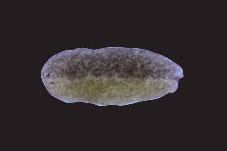
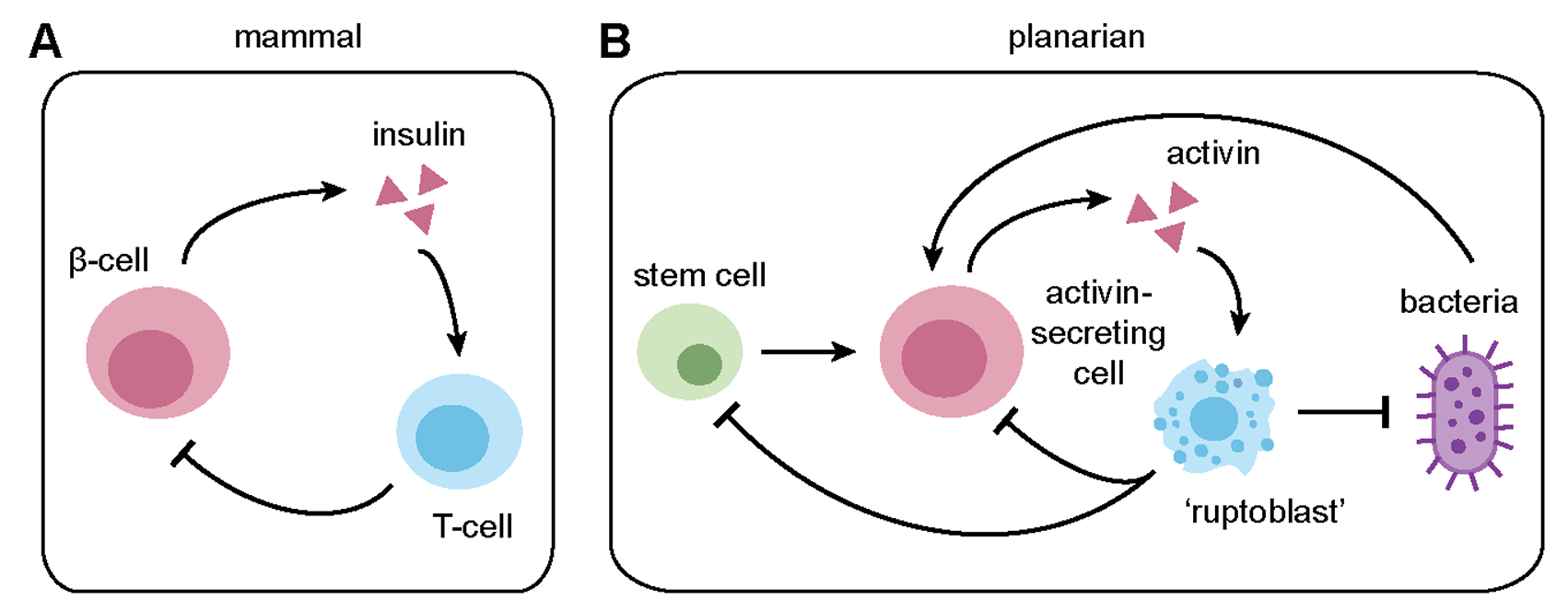
In planarian flatworms, a newly identified cell type appears to protect the body by bursting apart, releasing a lethal chemical blast that can kill nearby bacteria and damaged cells in minutes. The cells, called ruptoblasts, do not fit the usual picture of immune cells drawn from the classic white-blood-cell lineage. Instead, the work points to a very different branch of biology, one in which gland-like cells take on a fast, sacrificial defense role.
The study, published in Cell, was led by Prof. Benyamin Rosental of Ben-Gurion University of the Negev and Prof. Bo Wang of Stanford University.
What makes the finding stand out is not just the violence of the response, but the trigger. The cells react to activin, a hormone that helps regulate regeneration, reproduction, and normal tissue balance in the flatworm Schmidtea mediterranea. When activin rises too high, ruptoblasts answer with what the authors call “ruptosis,” a rapid, explosive death that destroys nearby targets in a tightly limited zone.
The team began with a puzzle. Much of what scientists know about immunity comes from a small set of model animals, yet the animal kingdom has many ways to solve the same problems, including clearing pathogens and recognizing cells that should not be there. The researchers wanted to know whether animals that lack adaptive immunity might have evolved other ways to achieve similar results.
In planarians, activin turned out to be more than a hormone. The researchers found that it also acts like an inflammatory signal. When they injected one form of planarian activin, called ACT-2, the animals showed increased activity in molecular pathways tied to inflammation. They also developed localized tissue damage and elevated cell death near the injection site.
That linked hormonal imbalance to immune activation. But it still did not explain which cells were doing the killing.
To find out, the team broke planarians into single cells and exposed them to ACT-2. A subset of cells suddenly lysed within about five minutes. Control proteins did not produce the same effect. Further analysis narrowed the reaction to one distinct population of highly granular cells. When activin was added, about 60 to 70 percent of those cells burst, spilling granules that quickly dispersed and vanished.
The researchers named the process ruptosis and the cells ruptoblasts.
That mechanism sets ruptoblasts apart from better-known immune tactics.
T cells and natural killer cells usually kill through direct contact. Neutrophils can release extracellular traps, but that process is different and often slower. Ruptoblasts did neither. They did not appear to touch their targets directly, and they did not release DNA traps. Instead, they seemed to discharge diffusive toxic agents into the surrounding space.
In imaging experiments, cells near activated ruptoblasts died soon after the burst. In the animal itself, ACT-2 injections wiped out ruptoblasts at the injection site, along with nearby activin-secreting cells and neoblasts, the stem cells that fuel planarian regeneration, within roughly 100 micrometers.
That matters because it suggests a way to eliminate both problem cells and the cells that might keep replenishing them.
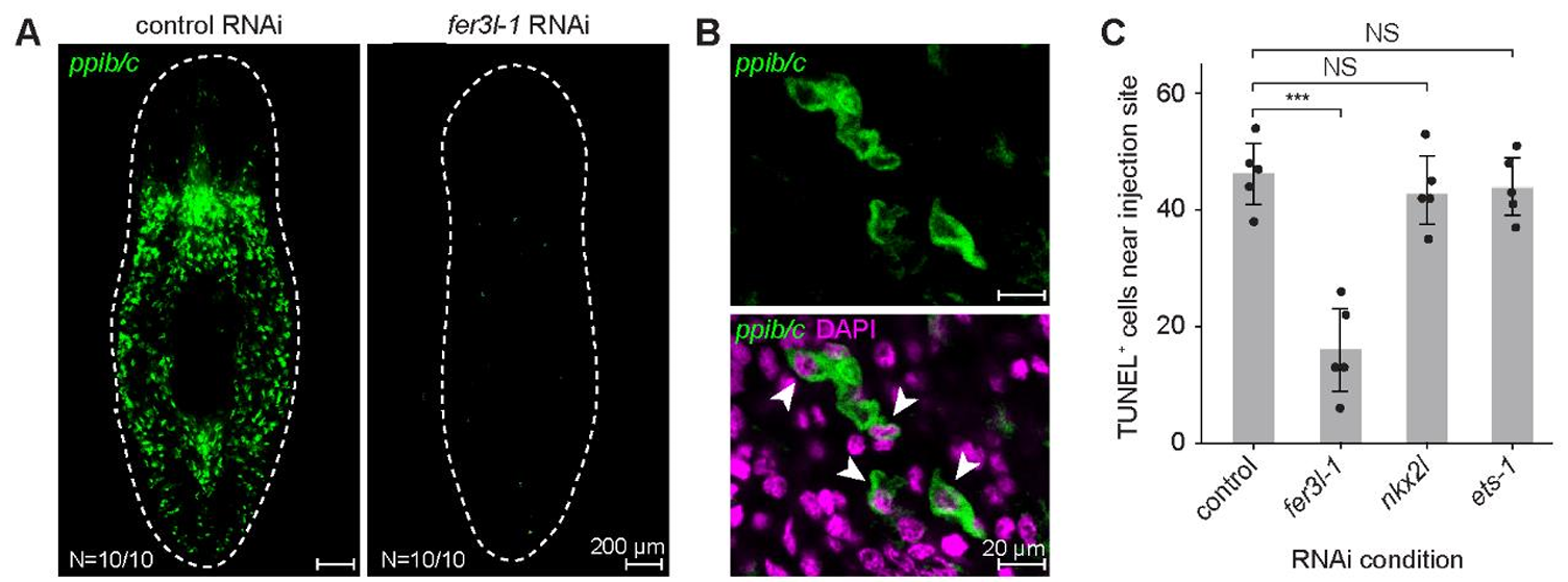
The work also showed that ruptoblasts are not the same as planarian cells previously suspected of handling many immune functions. Single-cell RNA sequencing and follow-up experiments tied the response specifically to one glandular cell cluster. When the researchers selectively removed that cluster, ACT-2-induced cell death and inflammatory signaling dropped sharply.
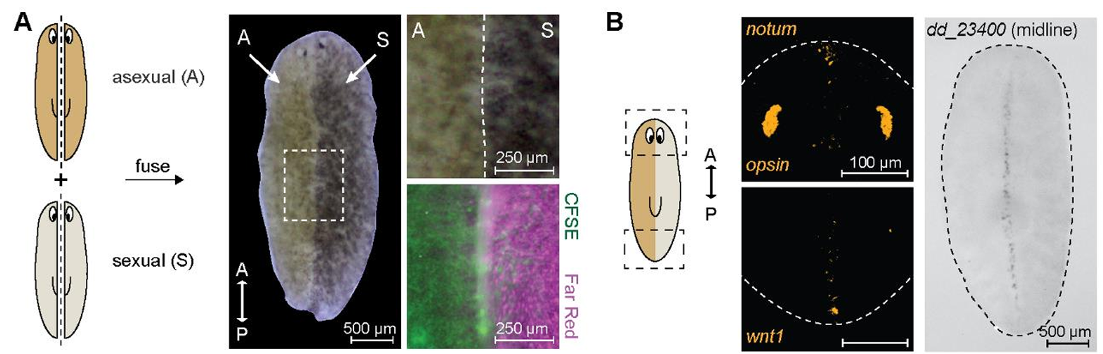
The study then moved from injections to a more dramatic test. The team fused asexual and sexual planarians together to create genetic chimeras, hoping to see whether mismatched tissues would provoke a rejection response.
They did.
The fused animals showed signs of chronic inflammation. The two genotypes stayed segregated instead of blending. The chimeras failed to feed, and about 40 percent developed lesions at the fusion site by around 14 days after fusion, followed by whole-body disintegration within a day.
Molecular measurements pointed again to elevated activin signaling and inflammation. When the researchers blocked a key part of that pathway, the lesions did not appear. When they removed ruptoblasts before fusion, lysis was prevented and inflammatory activity fell.
That result gave ruptoblasts a second clear role. They were not just involved in local responses to hormone spikes, but in a tissue-rejection process resembling a transplant conflict.
The same system also seemed to matter during infection.
When the researchers exposed planarians to pathogenic Pseudomonas, bacterial challenge activated the activin pathway, but only above a certain load. Animals in which activin signaling or ruptoblasts had been reduced became more sensitive to infection and lysed days earlier than controls. They also carried higher bacterial loads.
The next question was whether ruptoblasts could kill microbes directly.
On their own, the cells did not react to nearby bacteria. But when activin was added, they began to rupture within about five minutes, and nearby bacteria soon lost green fluorescent signal, a sign of membrane leakage, before taking up propidium iodide, which marks loss of membrane integrity. About 45 percent of the bacteria surrounding a ruptoblast were killed in a single event.
That suggests the cells do not patrol for microbes the way typical immune cells do. Instead, they may serve as a secondary weapon, switched on by signals from other cells that detect trouble first.
The authors suggest that cathepsin-positive phagocytic cells, which can engulf bacteria and also express activin, may help trigger ruptoblasts during infection.
The discovery also pushes at a deeper evolutionary question.
Ruptoblasts do not carry the standard immune markers scientists often use to identify defensive cells, and they seem to arise from a lineage distinct from classical immune cell types. Yet the researchers found similar gene-expression signatures in specific cell types across other flatworms, annelids, and acoel worms, animals that sit near the base of the bilaterian tree. Comparable cells appeared absent in cnidarians and in heavily studied systems such as vertebrates, flies, and nematodes.
That pattern hints that ruptoblast-like cells may be ancient, then lost in some major animal lineages.
It also helps explain why they escaped notice for so long. Modern biology leans heavily on standard lab models. If a cell type sits outside those systems and lacks familiar immune markers, it can remain effectively invisible even if versions of it are widespread elsewhere.
“This suggests that ruptoblasts may represent an ancient, bilaterian-specific immune cell type,” the authors wrote.
The work does not show that humans have ruptoblasts, and the study was done in planarian flatworms, not in mammals. But it offers a new example of how immune defense can be organized, one that links a hormone signal to a rapid, spatially restricted killing event.
That could matter in several ways. The released cocktail from ruptoblasts was potent enough to damage mammalian cells in lab tests, including human kidney cancer cells, which suggests that the underlying chemistry may have broad activity. The findings also point to a defense strategy that can strike multiple nearby targets at once, unlike slower contact-based killing. And because ruptosis is a one-time event, it may avoid some problems tied to prolonged immune activation.
The study leaves major questions open, including what molecules actually drive the burst, how the toxic agents work, and whether comparable cells in other animals use the same system. Even so, it expands the map of immunity in a way that could eventually inform efforts to design new cellular therapies or new methods for breaking apart bacterial communities.
Research findings are available online in the journal Cell.
The original story “Scientists discover ‘explosive’ new immune cell type” is published in The Brighter Side of News.
Like these kind of feel good stories? Get The Brighter Side of News’ newsletter.
The post Scientists discover ‘explosive’ new immune cell type appeared first on The Brighter Side of News.


Leave a comment
You must be logged in to post a comment.