DNA sits in sunlight every day, absorbing ultraviolet radiation that can set off the kind of chemical changes linked to mutations, aging, and cancer. Yet most of the time, the molecule avoids catastrophe.
New simulations suggest that this resilience depends not on one built-in escape route, but on a crowded, ultrafast network of molecular reactions. These reactions can dump harmful energy almost as soon as it appears.
The research focused on guanine and cytosine, two of DNA’s four chemical bases, arranged in short stacked segments that mimic key features of the double helix. By tracking their behavior at the atomic scale, the team found that once UV energy enters the system, the excited state quickly shifts into charge-transfer arrangements. Then, it relaxes through several competing pathways, many involving tightly linked movements of electrons and protons.
That picture is more complicated than the simpler models that have often dominated the field. Instead of following a single preferred track, the excited DNA fragments explored multiple routes back to stability. Often, this happened within femtoseconds. Each one of those routes adds to the molecule’s ability to avoid lasting injury.
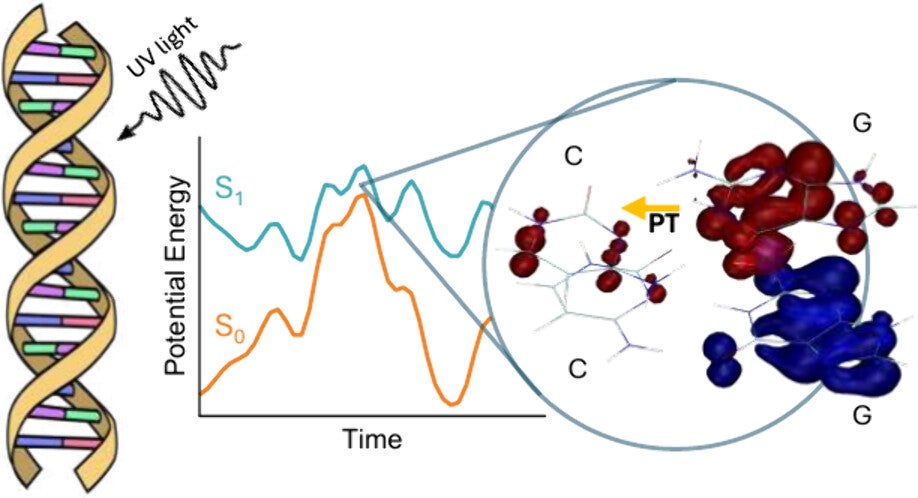
The study, led by researchers at the University of Surrey with collaborators at Aix Marseille University, the French National Centre for Scientific Research, and Université Claude Bernard Lyon 1, appeared in The Journal of Physical Chemistry Letters. The team used nonadiabatic dynamics simulations and high-level quantum chemical methods to examine two versions of a guanine-cytosine tetramer. One was alternating in sequence and one was nonalternating.
Those tiny systems were chosen because they preserve two features that matter in real DNA: hydrogen bonding between paired bases and stacking interactions between neighboring bases. Together, those interactions shape how excited states form and decay.
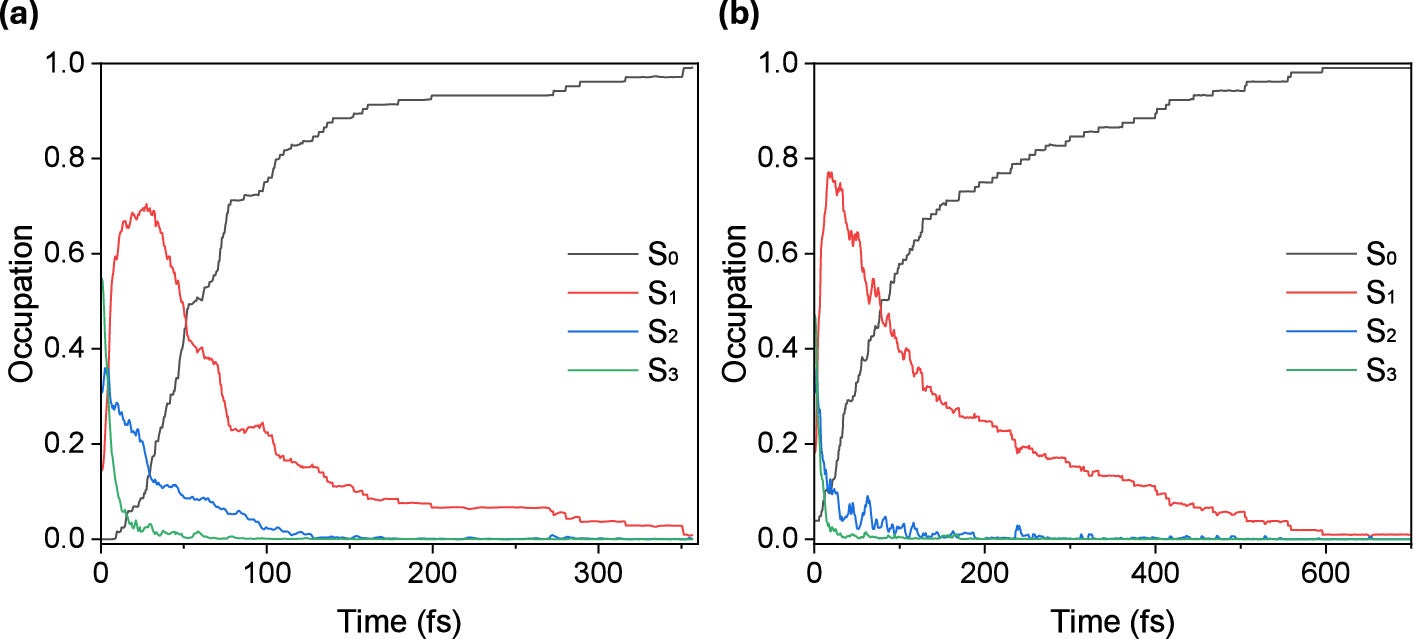
The simulations showed that soon after photoexcitation, the systems rapidly funneled toward a charge-transfer state in which electronic charge shifts from guanine to cytosine. In less than 50 femtoseconds, populations that began in higher excited states dropped into the lowest excited state. From there, decay to the ground state unfolded with average time constants of 64 femtoseconds in the nonalternating tetramer. In the alternating one, this decay took 141 femtoseconds.
Those times are strikingly short. A femtosecond is one quadrillionth of a second, a timescale so small that the protection happens long before ordinary biological damage could spread through the molecule.
Dr. Marco Sacchi, associate professor of physical and computational chemistry at the University of Surrey and senior author of the study, said: “DNA has evolved under constant exposure to UV radiation, yet it is extraordinarily resilient. What’s exciting about this research is that we can now see the incredibly fast molecular processes that safely drain away the energy before damage has a chance to spread. Understanding how Nature has developed these built-in defence mechanisms could help us better understand everything from mutation and ageing to the ways radiation affects living cells.”
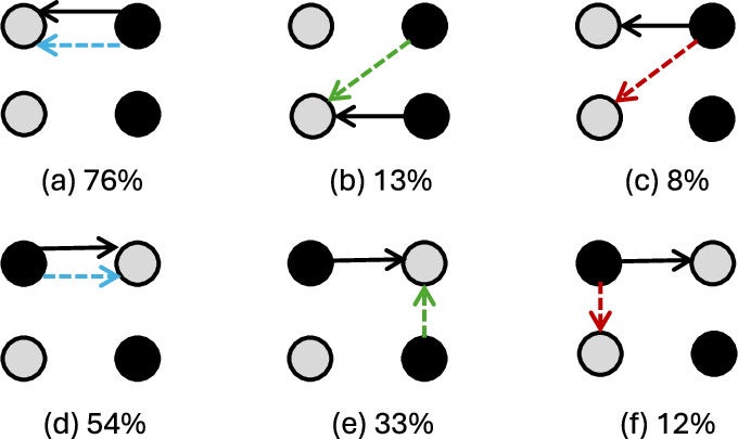
One of the central findings involved proton transfer between guanine and cytosine. The dominant route was a single proton transfer along the central hydrogen bond, from guanine to cytosine. That mechanism accounted for 82% of the relevant cases in the alternating tetramer and 70% in the nonalternating one.
A second pathway, involving transfer from a guanine amino group to a cytosine oxygen atom, also contributed. It appeared in 24% of trajectories in the nonalternating tetramer and 16% in the alternating one. Much rarer were transfers in the opposite direction, from cytosine to guanine.
What made the picture especially interesting was that proton transfer did not always line up neatly with electron transfer. Sometimes the charge moved between the same pair of bases involved in proton motion. In other cases, the electron shifted toward a different cytosine or along the same strand, especially in the alternating sequence. The result was a more flexible and dynamic process than a simple one-step handoff.
Juliana Gonçalves de Abrantes, postgraduate researcher at the University of Surrey and lead author, said: “What surprised us most was the diversity of the relaxation pathways, the different ways DNA can safely get rid of harmful UV energy. The electron and proton motions are strongly coupled, meaning they closely influence each other, but they are not rigidly locked together. This creates a rich network of possible decay routes that collectively enhance DNA photostability.”
That distinction matters because earlier debates in the field have often centered on which single mechanism dominates. The new work argues that the better answer may be that several mechanisms operate at once. The exact route may depend on sequence, geometry, and fleeting changes in electronic structure.
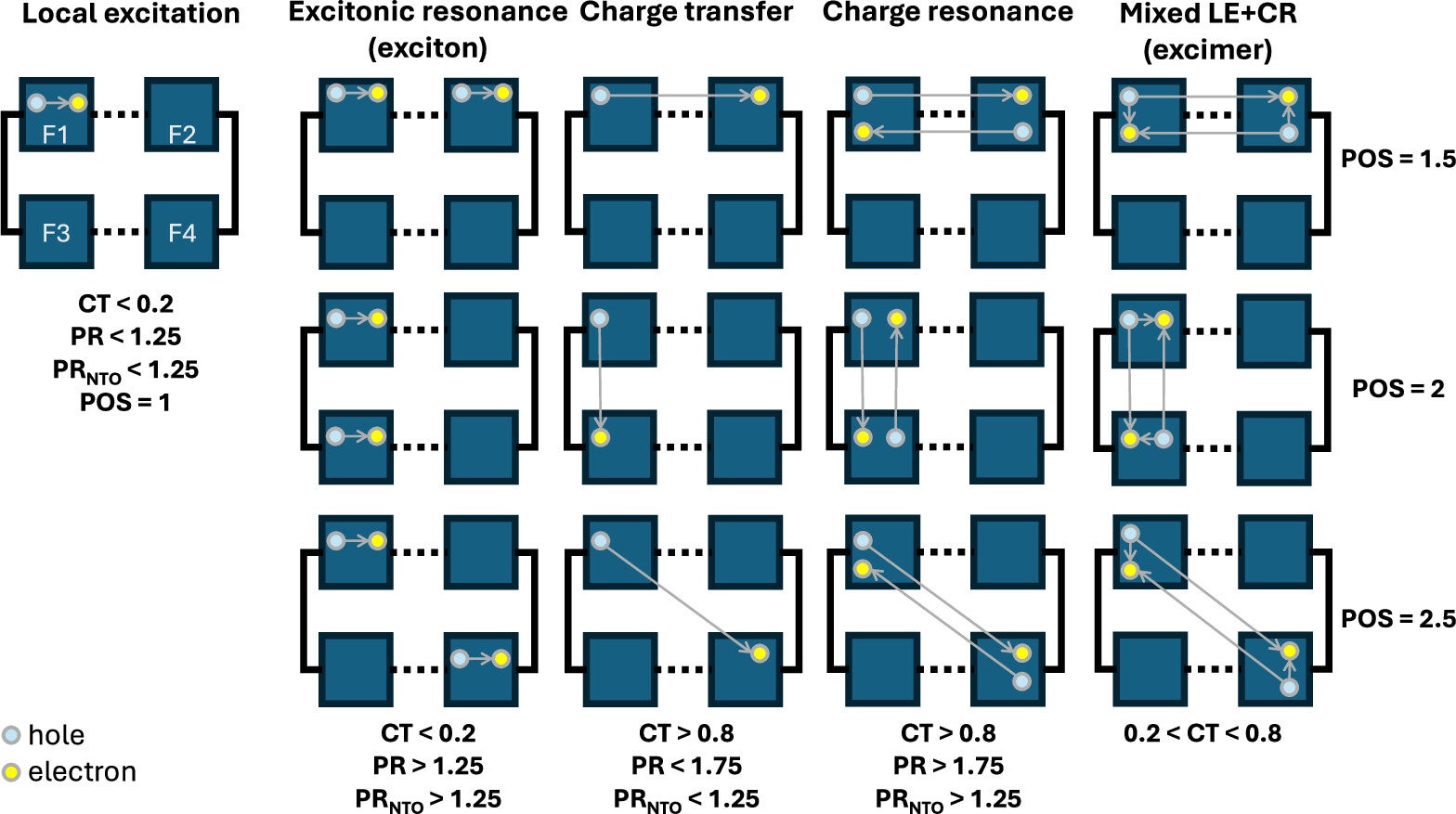
The team found that the photoexcited tetramers did not begin from one clean electronic state. Small distortions in structure and a dense cluster of nearly degenerate excited states meant the systems initially sampled a mix of local excitation, excitonic resonance, and mixed states. Even so, their later behavior converged. They quickly developed strong charge-transfer character before relaxing.
That common drift suggests charge separation is a key stage in the protective process, even if the next steps vary from one trajectory to another. In that sense, several ideas proposed in earlier studies may all be partly right. Each one describes one outcome within a wider ensemble of possible reactions.
The authors are careful not to oversell the result. Their mechanistic picture comes from a tetramer model in the gas phase and from a specific time-dependent density functional theory framework. They note that the balance between local excitation and charge-transfer states can depend on both the computational method and the starting geometry.
Additional work using solvent-inclusive QM/MM models could change how these pathways look in more realistic biological settings. Such work might help researchers test rarer outcomes such as cyclobutane pyrimidine dimer formation.
Still, the study gives a more unified real-time picture of how coupled charge and proton motions help DNA survive UV exposure. Rather than a single molecular trick, DNA seems to rely on a repertoire.
The findings sharpen the basic picture of how DNA withstands one of the oldest environmental threats on Earth: ultraviolet radiation from the Sun.
By showing that energy dissipation depends on several ultrafast, competing pathways rather than one dominant route, the study could help researchers think more clearly about when protection succeeds and when it fails.
That has relevance for cancer biology, because mutations often begin with DNA damage that escapes repair. It also matters for aging research, radiation biology, and biotechnology. In all those fields, understanding how genetic material responds to light can shape everything from damage models to new photoprotective strategies.
The work may also interest astrobiology, since DNA’s ability to survive intense radiation is tied to larger questions about how life persists under harsh conditions.
Research findings are available online in The Journal of Physical Chemistry Letters.
The original story “Researchers discover DNA’s hidden defense against UV radiation” is published in The Brighter Side of News.
Like these kind of feel good stories? Get The Brighter Side of News’ newsletter.
The post Researchers discover DNA’s hidden defense against UV radiation appeared first on The Brighter Side of News.