A severe burn changes life in an instant. Beyond the pain, it destroys the body’s natural shield against infection, dehydration and injury. For doctors, restoring that barrier quickly can mean the difference between recovery and tragedy. Yet despite decades of advances in medicine, rebuilding healthy skin remains one of the greatest challenges in wound care.
Now, researchers in Sweden have developed a promising new technology that could bring medicine closer to that goal. Scientists at the Center for Disaster Medicine and Traumatology and Linköping University have created what they describe as a form of “skin in a syringe,” a living gel packed with cells that can be injected or even 3D printed into skin grafts. Their findings suggest the material could help the body rebuild functional skin rather than forming scar tissue.
The innovation arrives at a time when the need for better wound treatments is growing. Burns are the world’s fourth most common form of trauma. About 11 million people seek medical care for burn injuries every year, and roughly 180,000 die annually. Chronic wounds also place a massive burden on healthcare systems. In the United States alone, Medicare spends an estimated $96.8 billion each year on wound care.
Researchers believe their new approach could eventually offer a better path forward.
Healthy skin is far more complex than it appears. The outer layer, known as the epidermis, acts as a protective barrier. Beneath it lies the dermis, a thicker and far more sophisticated layer containing blood vessels, nerves, hair follicles and connective tissue that gives skin its strength and flexibility.
Current treatments often focus on replacing the epidermis. Large burns are commonly treated using split-thickness skin grafts, which transplant the outer layer and a thin portion of the dermis from another part of the patient’s body.
While these procedures save lives, they often leave significant scarring. The deeper dermal layer rarely regenerates completely.
“The dermis is so complicated that we can’t grow it in a lab. We don’t even know what all its components are. That’s why we, and many others, think that we could possibly transplant the building blocks and then let the body make the dermis itself,” said Johan Junker, researcher at the Swedish Center for Disaster Medicine and Traumatology and docent in plastic surgery at Linköping University.
Creating those building blocks became the central challenge for the research team.
The scientists focused on fibroblasts, the most common cells found in the dermis. These connective tissue cells produce collagen, elastin and other materials that give skin its structure. They also help coordinate healing and can develop into more specialized cell types when needed.
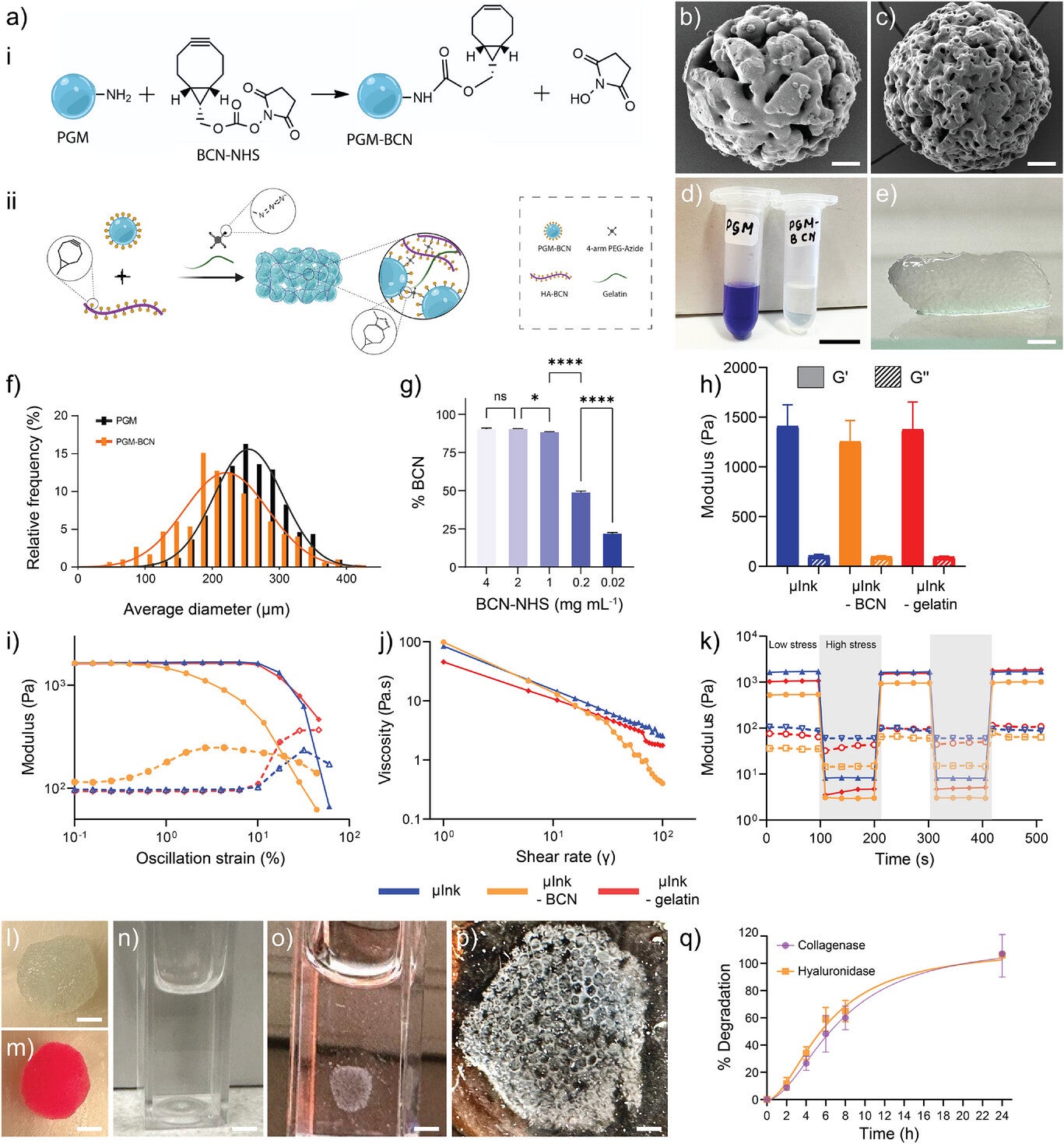
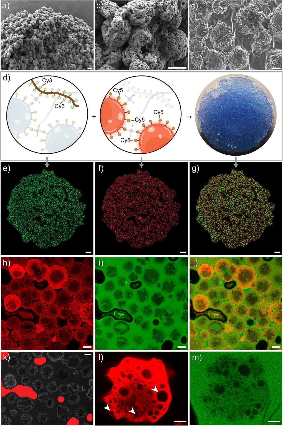
Fibroblasts are relatively easy to collect and grow in the laboratory. The researchers expanded these cells on tiny porous gelatin particles known as microcarriers. Made from collagen-like material, these sponge-shaped particles provide a scaffold where cells can attach, multiply and begin producing new tissue.
Within days, the fibroblasts spread across the microcarriers and multiplied rapidly. The cells remained active and began producing many of the proteins normally found in healthy dermis.
The challenge was keeping these cell-loaded particles in place after transplantation.
A simple liquid would quickly flow away from a wound. The team needed something that could hold the cells together while remaining easy to apply.
The solution combined the gelatin particles with hyaluronic acid, a naturally occurring substance found throughout the body. Using a process called click chemistry, the researchers linked the components together to create a stable yet flexible gel.
The result behaves in a remarkable way.
“The gel has a special feature that means that it becomes liquid when exposed to light pressure. You can use a syringe to apply it to a wound, for example, and once applied it becomes gel-like again. This also makes it possible to 3D print the gel with the cells in it,” said Daniel Aili, professor of molecular physics at Linköping University.
Scientists call this behavior shear-thinning. Under pressure, the material flows like a liquid. Once the pressure disappears, it quickly returns to a solid-like state.
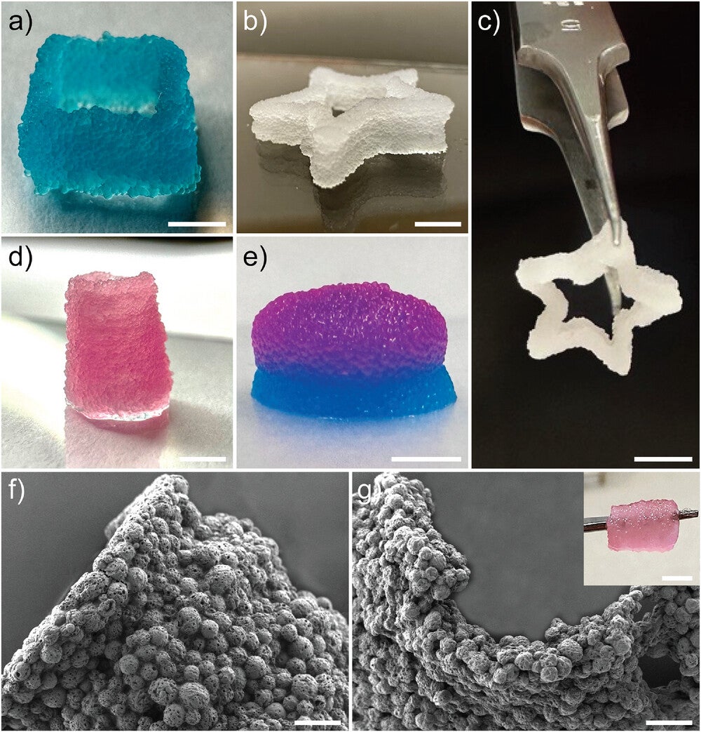
This property allows the gel to be injected directly into wounds or printed into complex three-dimensional structures.
The researchers successfully printed cylinders, star-shaped structures and multilayer constructs that remained stable for more than 45 days in laboratory conditions.
The material does more than simply hold cells together.
The porous structure allows oxygen and nutrients to reach the cells. At the same time, the gel gradually breaks down as natural healing progresses.
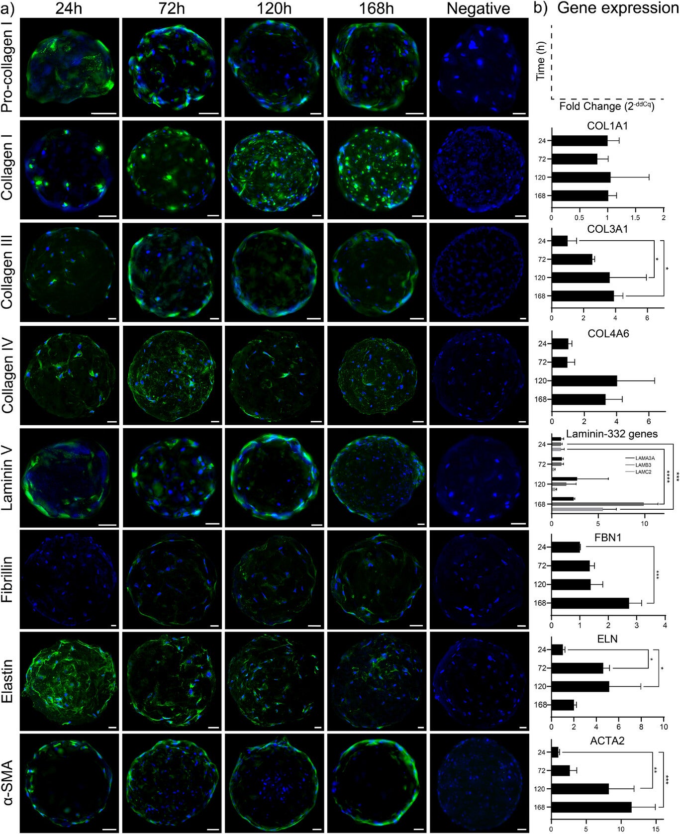
Tests showed that the fibroblasts remained highly viable after printing. More importantly, they continued producing key proteins needed for healthy skin formation.
Levels of collagen I, collagen III, collagen IV, laminin and fibrillin all increased over time. These proteins help create the supportive framework that gives skin its strength, flexibility and resilience.
Researchers also observed signs that the cells were actively remodeling their environment. Rather than creating disorganized scar tissue, they appeared to be building a structure that more closely resembled normal dermis.
This distinction is critical because scar tissue lacks many of the functions of healthy skin. It often appears stiff, weak and less elastic than surrounding tissue.
To see how the material performed inside the body, the team implanted small 3D-printed constructs beneath the skin of mice.
The results were encouraging.
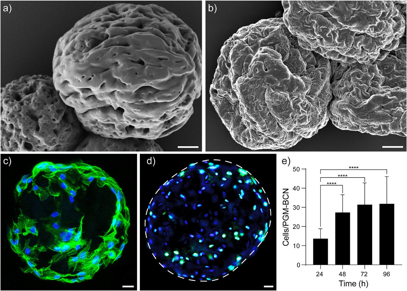
The implanted cells survived, continued growing and remained biologically active for at least 28 days. Researchers observed increasing collagen production throughout the study period.
Most importantly, blood vessels formed within the implants.
“We see that the cells survive and it’s clear that they produce different substances that are needed to create new dermis. In addition, blood vessels are formed in the grafts, which is important for the tissue to survive in the body. We find this material very promising,” Junker said.
Blood vessel formation represents one of the biggest obstacles in tissue engineering. Without a blood supply, cells cannot receive enough oxygen and nutrients to survive long term.
The presence of new blood vessels suggests the material may integrate successfully with living tissue.
The implications may extend far beyond wound healing.
Scientists have long struggled to create larger engineered tissues and organoids because cells in the center often die from lack of oxygen. Blood vessel networks remain one of the major bottlenecks limiting the growth of artificial tissues.
In a related study, the Linköping University team developed hydrogel threads that contain about 98% water. These threads can be tied into knots, formed into miniature tubes and used to transport fluids.
“The hydrogel threads become quite elastic, so we can tie knots on them. We also show that they can be formed into mini-tubes, which we can pump fluid through or have blood vessel cells grow in,” Aili said.
These tiny channels may eventually help create artificial blood vessel networks inside engineered tissues and organoids.
Although the research remains in the experimental stage, the findings point toward a future where doctors could take a small skin sample from a patient, grow the cells in the laboratory and create personalized grafts using 3D printing.
Instead of simply closing wounds, future treatments might help regenerate functional skin that behaves more like the original tissue.
The researchers plan additional studies, including testing in larger animal models that more closely resemble human wound healing.
Much work remains before the technology reaches hospitals. Yet the early results suggest that combining living cells, advanced biomaterials and 3D printing could open a new chapter in regenerative medicine.
This technology could significantly improve treatment options for patients with severe burns, chronic wounds and traumatic injuries. By promoting the formation of functional dermal tissue instead of scar tissue, it may improve long-term mobility, appearance and quality of life for patients recovering from major skin damage.
The findings also address one of tissue engineering’s most persistent challenges: creating structures that develop their own blood supply. The observed formation of blood vessels inside the grafts suggests the material may support larger and more complex engineered tissues in the future.
Beyond wound care, the technology could contribute to advances in regenerative medicine, organoid development and artificial tissue production. Researchers may eventually use similar materials to build more realistic laboratory models for studying disease, testing drugs and developing future organ replacement therapies. By helping cells survive, organize and integrate within the body, this approach moves science closer to repairing damaged tissues rather than simply replacing them.
Research findings are available online in the journal Advanced Healthcare Materials.
The original story “Scientists 3D print living skin that helps grow blood vessels” is published in The Brighter Side of News.
Like these kind of feel good stories? Get The Brighter Side of News’ newsletter.
The post Scientists 3D print living skin that helps grow blood vessels appeared first on The Brighter Side of News.

